2010
MRI follow-up of conservatively treated meniscal knee lesions in general practice
Publication
Publication
European Radiology: journal of the European Congress of Radiology , Volume 20 - Issue 5 p. 1242- 1250
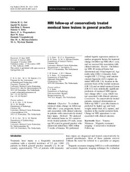
Objective: To evaluate meniscal status change on follow-up MRI after 1 year, prognostic factors and association with clinical outcome in patients with conservatively treated knee injury. Methods: We analysed 403 meniscal horns in 101 conservatively treated patients (59 male; mean age 40 years) in general practice who underwent initial knee MRI within 5 weeks of trauma. We performed ordinal logistic regression analysis to analyse prognostic factors for meniscal change on follow-up MRI after 1 year, and we assessed the association with clinical outcome. Results: On follow-up MRI 49 meniscal horns had deteriorated and 18 had improved. Age (odds ratio [OR] 1.3/decade), body weight (OR 1.2/10 kg), total anterior cruciate ligament (ACL) rupture on initial MRI (OR 2.4), location in the posterior horn of the medial meniscus (OR 3.0) and an initial meniscal lesion (OR 0.3) were statistically significant predictors of meniscal MRI appearance change after 1 year, which was not associated with clinical outcome. Conclusion: In conservatively treated patients, meniscal deterioration on follow-up MRI 1 year after trauma is predicted by higher age and body weight, initial total ACL rupture, and location in the medial posterior horn. Change in MRI appearance is not associated with clinical outcome.
| Additional Metadata | |
|---|---|
| , , , , , , , , , , , , , , , , , , , , , , , , , , , , , , , , , , | |
| doi.org/10.1007/s00330-009-1648-3, hdl.handle.net/1765/19811 | |
| European Radiology: journal of the European Congress of Radiology | |
| Organisation | Erasmus MC: University Medical Center Rotterdam |
|
Oei, E., Koster, I., Hensen, J. H. J., Boks, S., Wagemakers, H., Koes, B., … Hunink, M. (2010). MRI follow-up of conservatively treated meniscal knee lesions in general practice. European Radiology: journal of the European Congress of Radiology, 20(5), 1242–1250. doi:10.1007/s00330-009-1648-3 |
|