1996
GATA transcription factors associate with a novel class of nuclear bodies in erythroblasts and megakaryocytes.
Publication
Publication
EMBO Journal , Volume 15 p. 319- 333
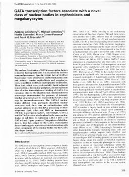
The nuclear distribution of GATA transcription factors in murine haemopoietic cells was examined by indirect immunofluorescence. Specific bright foci of GATA-1 fluorescence were observed in erythroleukaemia cells and primary murine erythroblasts and megakaryocytes, in addition to diffuse nucleoplasmic localization. These foci, which were preferentially found adjacent to nucleoli or at the nuclear periphery, did not represent sites of active transcription or binding of GATA-1 to consensus sites in the beta-globin loci. Immunoelectron microscopy demonstrated the presence of intensely labelled structures likely to represent the GATA-1 foci seen by immunofluorescence. The GATA-1 nuclear bodies differed from previously described nuclear structures and there was no co-localization with nuclear antigens involved in RNA processing or other ubiquitous (Spl, c-Jun and TBP) or haemopoietic (NF-E2) transcription factors. Interestingly, GATA-2 and GATA-3 proteins also localized to the same nuclear bodies in cell lines co-expressing GATA-1 and -2 or GATA-1 and -3 gene products. This pattern of distribution is, thus far, unique to the GATA transcription factors and suggests a protein-protein interaction with other components of the nuclear bodies via the GATA zinc finger domain.
| Additional Metadata | |
|---|---|
| , , , , , , , , , , , , , , , , , , , , , , , , , , , , , , , , | |
| hdl.handle.net/1765/2521 | |
| EMBO Journal | |
| Organisation | Erasmus MC: University Medical Center Rotterdam |
|
Elefanty, A., Antoniou, M., Custodio, N., Carmo-Fonseca, M., & Grosveld, F. (1996). GATA transcription factors associate with a novel class of nuclear bodies in erythroblasts and megakaryocytes. EMBO Journal, 15, 319–333. Retrieved from http://hdl.handle.net/1765/2521 |
|