1974
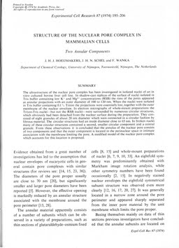
Structure of the nuclear pore complex in mammalian cells. Two annular components.
Publication
Publication
Experimental Cell Research: emphasizing molecular approaches to cell biology , Volume 87 - Issue 1 p. 195- 206
The ultrastructure of the nuclear pore complex has been investigated in isolated nuclei of an in vitro cultured bovine liver cell line. In shadow-cast replicas of the surface of nuclei isolated in Tris buffer containing low K+ and Mg2+ concentrations (RSB) the rims of the pores appeared as annular projections with an outer diameter of 100 to 120 nm. When the nuclei were isolated in Tris buffer containing 0.1% Triton the projections were essentially lost, together with the outer membrane of the nuclear envelope. In electron micrographs of whole-mount preparations the Triton-Tris nuclei—but not the RSB nuclei—were surrounded by numerous circular structures, which obviously had been detached from the nuclear surface during the preparation. They consisted of eight granules of about 20 nm diameter which were connected in a circular fashion by fibrous material. The circular structures had an inside diameter close to 65 nm. In broken nuclei many of these circular structures contained a second, smaller circular component and a central granule. From these observations it is concluded that the annulus of the nuclear pore consists of two components and that the outer component is located in the perinuclear space in intimate association with the membrane limiting the pore. A modified model of the nuclear pore complex which accounts for this location is proposed.
| Additional Metadata | |
|---|---|
| , , , , , , , , , , , , , , , , , , , , , , | |
| doi.org/10.1016/0014-4827(74)90542-4, hdl.handle.net/1765/2941 | |
| Experimental Cell Research: emphasizing molecular approaches to cell biology | |
| Organisation | Erasmus MC: University Medical Center Rotterdam |
|
Hoeijmakers, J., Schel, J. H. N., & Wanka, F. (1974). Structure of the nuclear pore complex in mammalian cells. Two annular components. Experimental Cell Research: emphasizing molecular approaches to cell biology, 87(1), 195–206. doi:10.1016/0014-4827(74)90542-4 |
|