1993
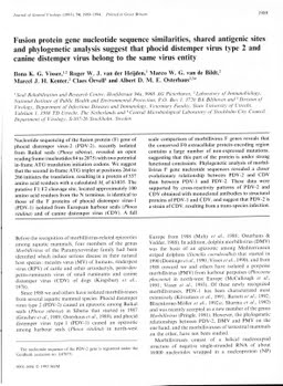
Fusion protein gene nucleotide sequence similarities, shared antigenic sites and phylogenetic analysis suggest that phocid distemper virus 2 and canine distemper virus belong to the same virus entity.
Publication
Publication
Journal of General Virology , Volume 74 p. 1989- 1994
Nucleotide sequencing of the fusion protein (F) gene of phocid distemper virus-2 (PDV-2), recently isolated from Baikal seals (Phoca sibirica), revealed an open reading frame (nucleotides 84 to 2075) with two potential in-frame ATG translation initiation codons. We suggest that the second in-frame ATG triplet at positions 264 to 266 initiates the translation, resulting in a protein of 537 amino acid residues with a calculated M(r) of 63,035. The putative F1/F2 cleavage site, located approximately 100 amino acid residues from the N terminus, is identical to those of the F proteins of phocid distemper virus-1 (PDV-1) isolated from European harbour seals (Phoca vitulina) and of canine distemper virus (CDV). A full scale comparison of morbillivirus F genes reveals that the conserved F0 extracellular protein-encoding region contains a large number of non-expressed mutations, suggesting that this part of the protein is under strong functional constraints. Phylogenetic analysis of morbillivirus F gene nucleotide sequences revealed a closer evolutionary relationship between PDV-2 and CDV than between PDV-1 and PDV-2. These data were supported by cross-reactivity patterns of PDV-2 and CDV obtained with monoclonal antibodies to structural proteins of PDV-1 and CDV, and suggest that PDV-2 is a strain of CDV, resulting from a trans-species infection.
| Additional Metadata | |
|---|---|
| , , , , , , , , , , , , , , , , , , , , , | |
| hdl.handle.net/1765/3480 | |
| Journal of General Virology | |
| Organisation | Erasmus MC: University Medical Center Rotterdam |
|
Visser, I., van der Heijden, R., van de Bildt, M., Kenter, M., Örvell, C., & Osterhaus, A. (1993). Fusion protein gene nucleotide sequence similarities, shared antigenic sites and phylogenetic analysis suggest that phocid distemper virus 2 and canine distemper virus belong to the same virus entity. Journal of General Virology, 74, 1989–1994. Retrieved from http://hdl.handle.net/1765/3480 |
|