
2013-12-11
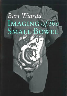
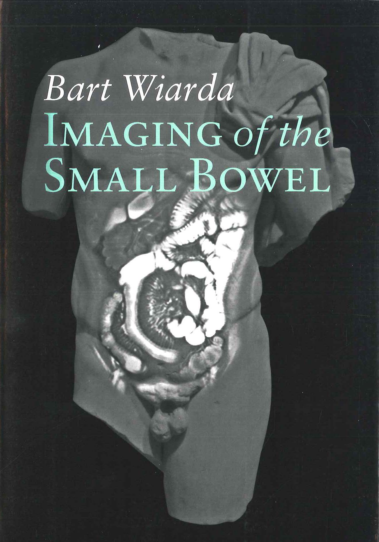
Imaging of the Small Bowel
Publication
Publication
Beeldvorming van de dunne darm
Abstract
Visualization of the small bowel is a clinical challenge due to its length, motility, shape, and central location distal to the stomach and proximal to the coecum. During the last decade, several new techniques have been introduced for visualizing the small bowel. Radiologists and clinicians are now faced with a dilemma in deciding which technique to use in two common groups of adult patients: those with known or suspected Crohn’s disease, and those with obscure gastrointestinal bleeding. This decision is primarily based on the accuracy, burden, and patient preference of each technique. In patients with suspected Crohn’s disease, diagnosing the disease and establishing the location, extent, and severity of the disease is essential. Symptoms of Crohn’s disease can sometimes overlap those of other common gastrointestinal problems, such as irritable bowel syndrome (IBS), celiac disease, and other gastrointestinal abnormalities. Crohn’s disease is characterized as a chronic, relapsing, and remitting inflammatory bowel disease (IBD), which can start at an early age and may require lifelong surveillance. In the last few decades the incidence of Crohn’s disease has continued to increase worldwide, and the prevalence and incidence are the same in Europe and the United States. A north-south gradient and lower incidence of Crohn’s disease is present in Asian and African people. Crohn’s disease has a predilection for the terminal ileum; the disease localizes to the small bowel in 70% of patients, but it can also occur in any other part of the gastrointestinal tract. The inflammation is characterized by patchy, transmural, granulomatous lesions, which can cause a range of complications, such as small bowel stenosis, fistulas, and abscesses. In addition, colon involvement increases the risk of developing colorectal cancer.
| Additional Metadata | |
|---|---|
| , , , , | |
| The COMRADE study (Chapter 6-8) was financially supported by • Foreest Medical School Netherlands • Bayer Germany • Siemens Netherlands The printing of this thesis was financially supported by • Clarity Translations • ABBOTT Immunology • Esaote Benelux • Foreest Medical School Netherlands • FUJIFILM Medical Systems Benelux • Holland Health • Oldelft Benelux B.V. • Toshiba Medical Systems Nederland • Tromp Medical BV • Vrest Medical | |
| J. Stoker (Jacob) , E.J. Kuipers (Ernst) | |
| Erasmus University Rotterdam | |
| hdl.handle.net/1765/37639 | |
| Organisation | Erasmus MC: University Medical Center Rotterdam |
|
Wiarda, B. (2013, December 11). Imaging of the Small Bowel. Retrieved from http://hdl.handle.net/1765/37639 |
|