
2013-10-23
Biomechanical Modeling of Atherosclerotic Plaques for Risk Assessment
Publication
Publication
Biomechanische Modelvorming van Atherosclerotische Plaques ten behoeve van Risico Analyse
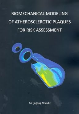
A healthy arterial wall comprises three layers: the adventitia, the media and the intima (Figure 1.1, left side). The adventitia is the outermost layer, mainly composed of collagen. The media underlies the adventitia and is the middle layer in the arterial wall. It is made up of concentrically arranged smooth muscle cells and collagen fibers. The intima is the innermost layer. It is a thin sheet of endothelial cells attached to a basal membrane. Atherosclerosis is a systemic, inflammatory disease of the arterial system characterized by local thickening of vessel walls. Thickened arterial segments are called atherosclerotic plaques (Figure 1.1, right side). During atherogenesis - progression of an atherosclerotic plaque- the major changes take place in the intima due to infiltration of lipids and inflammatory cells from the luminal side, smooth muscle cell migration and proliferation, extracellular matrix deposition, and intraplaque hemorrhage. From a thin cell layer, the intima transforms into a thick layer (Figure 1.1) with the possible structural components being smooth muscle cells, collagen and elastin fibers, and lipids. Besides changes in the intima, atherosclerosis causes differentiation in the media and adventitia layers. Fibrosis, atrophy and inflammation may take place in the media and adventitia during atherogenesis.
| Additional Metadata | |
|---|---|
| , | |
| This research project was funded by Medical Delta. Additional financial support by the Hamamatsu Photonics Deutschland GmbH, Cardialysis BV and FUJIFILM VisualSonics are gratefully acknowledged. | |
| A.F.W. van der Steen (Ton) | |
| Erasmus University Rotterdam | |
| hdl.handle.net/1765/41642 | |
| Organisation | Erasmus MC: University Medical Center Rotterdam |
|
Akyildiz, A. (2013, October 23). Biomechanical Modeling of Atherosclerotic Plaques for Risk Assessment. Retrieved from http://hdl.handle.net/1765/41642 |
|