1993
Digital geometric measurements in comparison to cinefilm analysis of coronary artery dimensions
Publication
Publication
Catheterization and Cardiovascular Interventions , Volume 28 p. 283- 290
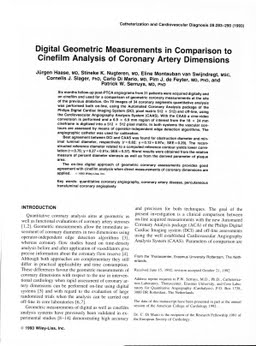
Six months follow-up post-PTCA angiograms from 31 patients were acquired digitally and on cinefilm and used for a comparison of geometric coronary measurements at the site of the previous dilatation. On 70 images of 34 coronary segments quantitative analysis was performed both on-line, using the Automated Coronary Analysis package of the Philips Digital Cardiac Imaging System (DCI, pixel matrix 512 x 512) and off-line, using the Cardiovascular Angiography Analysis System (CAAS). With the CAAS a cine-video conversion is performed and a 6.9 x 6.9 mm region of interest from the 18 x 24 mm cineframe is digitized into a 512 x 512 pixel matrix. In both systems the vascular contours are assessed by means of operator-independent edge detection algorithms. The angiographic catheter was used for calibration. Best agreement between DCI and CAAS was found for obstruction diameter and minimal luminal diameter, respectively (r = 0.82; y = 0.12 + 0.97x; SEE = 0.29). The reconstructed reference diameter related to a computed reference contour yields lower correlation (r = 0.76; y = 0.27 + 0.91x; SEE = 0.37). Worst results were obtained from the relative measure of percent diameter stenosis as well as from the derived parameter of plaque area. The on-line digital approach of geometric coronary assessments provides good agreement with cinefilm analysis when direct measurements of coronary dimensions are applied.
| Additional Metadata | |
|---|---|
| , , , , , , , , , , , , | |
| hdl.handle.net/1765/4501 | |
| Catheterization and Cardiovascular Interventions | |
| Organisation | Erasmus MC: University Medical Center Rotterdam |
|
Haase, J., Nugteren, S. K., Montauban van Swijndregt, E., Slager, C., di Mario, C., de Feyter, P., & Serruys, P. (1993). Digital geometric measurements in comparison to cinefilm analysis of coronary artery dimensions. Catheterization and Cardiovascular Interventions, 28, 283–290. Retrieved from http://hdl.handle.net/1765/4501 |
|