1993
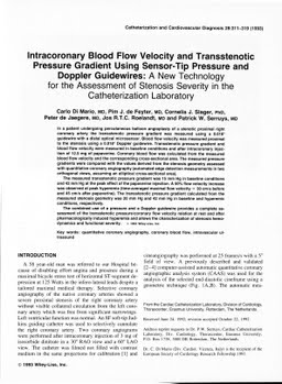
Intracoronary blood flow velocity and transstenotic pressure gradient using sensor-tip pressure and doppler guidewires: a new technology for the assessment of stenosis severity in the Catheterization Laboratory
Publication
Publication
Catheterization and Cardiovascular Interventions , Volume 28 p. 311- 319
In a patient undergoing percutaneous balloon angioplasty of a stenotic proximal right coronary artery the transstenotic pressure gradient was measured using a 0.018" guidewire with a distal optical microsensor. Blood flow velocity was measured proximal to the stenosis using a 0.018" Doppler guidewire. Transstenotic pressure gradient and blood flow velocity were measured in baseline conditions and after intracoronary injection of 12.5 mg of papaverine. Coronary blood flow was calculated from the measured blood flow velocity and the corresponding cross-sectional area. The measured pressure gradients were compared with the values derived from the stenosis geometry assessed with quantitative coronary angiography (automated edge detection measurements in two orthogonal views, assuming an elliptical cross-sectional area). The measured transstenotic pressure gradient was 15 mm Hg in baseline conditions and 42 mm Hg at the peak effect of the papaverine injection. A 50% flow velocity increase was observed at peak hyperemia (time-averaged maximal flow velocity = 30 cm/s before and 45 cm/s after papaverine). The transstenotic pressure gradient calculated from the measured stenosis geometry was 20 mm Hg and 42 mm Hg in baseline and hyperemic conditions, respectively. The combined use of a pressure and a Doppler guidewire provides a complete assessment of the transstenotic pressure/coronary flow velocity relation at rest and after pharmacologically induced hyperemia and allows the characterization of stenosis hemodynamics and functional severity.
| Additional Metadata | |
|---|---|
| , , , , , , , , , , , , , , , | |
| hdl.handle.net/1765/4502 | |
| Catheterization and Cardiovascular Interventions | |
| Organisation | Erasmus MC: University Medical Center Rotterdam |
|
di Mario, C., de Feyter, P., Slager, C., de Jaegere, P., Roelandt, J., & Serruys, P. (1993). Intracoronary blood flow velocity and transstenotic pressure gradient using sensor-tip pressure and doppler guidewires: a new technology for the assessment of stenosis severity in the Catheterization Laboratory. Catheterization and Cardiovascular Interventions, 28, 311–319. Retrieved from http://hdl.handle.net/1765/4502 |
|