
1990-11-21
Ascending and descending axon-collaterals from cervical spinal neurons : a retrograde fluorescent double-labeling study in the rat.
Publication
Publication
Opstijgende en afdalende axoncollateralen van neuronen gelegen in het cervicale ruggemerg : een onderzoek in de rat met behulp van de rettograde fluorescente dubbel-labeling methode
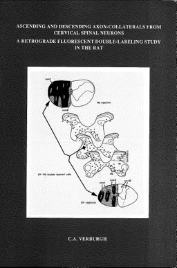
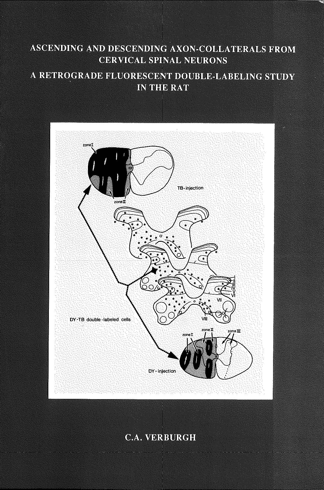
In order to gain insight into the function of the central nervous system it is of importance to know the lay-out of fiber connections between its constituent cells and cell-groups. Much is already known of the anatomy of the spinal cord and its afferent and efferent fibers; this will be reviewed in Chapter 2. A large part of the spinal neuronal network consists of propriospinal connections representing the anatomical basis for integration of information within the spinal cord itself. The present study was undertaken to investigate propriospinal neurons in more detail. Specifically, a search was made for neurons which project to more than one target area by way of divergent side-branches, or collaterals, of their axon. Earlier anatomical findings already suggested that such "branching" neurons' might be present within the spinal cord. Thus retrograde HRP studies in the cat [254, 286, 288, 359, 416] identified the spinal neurons which give rise to descending propriospinal fibers. The distribution of these neurons largely overlaps that of the neurons giving rise to ascending propriospinal and supraspinal fibers [286, 288]. Therefore some of these neurons may be "branching" neurons which give rise to ascending and descending collaterals. In fact, such neurons were already noted in several Golgi studies [68, 242, 344], but their collaterals could be traced only over short distances. Electrophysiological studies [9, 146, 173, 360] supported these Golgi findings, indicating that some cervical spinal neurons projecting to lumbar levels also give rise to ascending supraspinal collaterals. In the present series of experiments in the rat, an attempt was made: a) to determine the location and relative numbers of such branching neurons in the cervical cord, and b) to approximate the rostral and caudal extent of projection of their collaterals. To this purpose we used the retrograde fluorescent double-labeling method (described in Chapter 3). The results of the first set of experiments (Chapter 4), in which tracer injections were made at various spinal levels, indicated that many neurons in the cervical grey matter in the rat are branching neurons, distributing ascending collaterals to upper cervical or supraspinal levels, and descending collaterals to thoracic or lumbosacral levels. Most descending collaterals of the branching cervical neurons were found to terminate at short distances, i.e. in the upper thoracic cord. On the other hand, the majority of the ascending collaterals were distributed to supraspinal levels. Although the findings suggested that many of these collaterals projected to the caudal brainstem, it remained unsettled whether the supraspinally ascending fibers specifically belonged to any of the various ascending tracts. Therefore, further sets of experiments were designed to clarify this issue, investigating the presence and relative numbers of supraspinal collaterals to the cerebellum (Chapter 5), the thalamus and tectum (Chapter 6) and the dorsal medulla (Chapter 7). Summarizing, the results of the latter series of experiments indicate that relatively many of the supraspinally ascending collaterals are distributed to the medulla, only few to the thalamus and tectum and extremely few to the cerebellum.
| Additional Metadata | |
|---|---|
| , , , , | |
| Erasmus University Rotterdam | |
| J. Voogd (Jan) | |
| hdl.handle.net/1765/50789 | |
| Organisation | Erasmus MC: University Medical Center Rotterdam |
|
Verburgh, C. A. (1990, November 21). Ascending and descending axon-collaterals from cervical spinal neurons : a retrograde fluorescent double-labeling study in the rat.. Retrieved from http://hdl.handle.net/1765/50789 |
|