
1990-06-20
Human T cell differentiation : basic aspects and their clinical applications
Publication
Publication
T-cel differentiatie bij mens
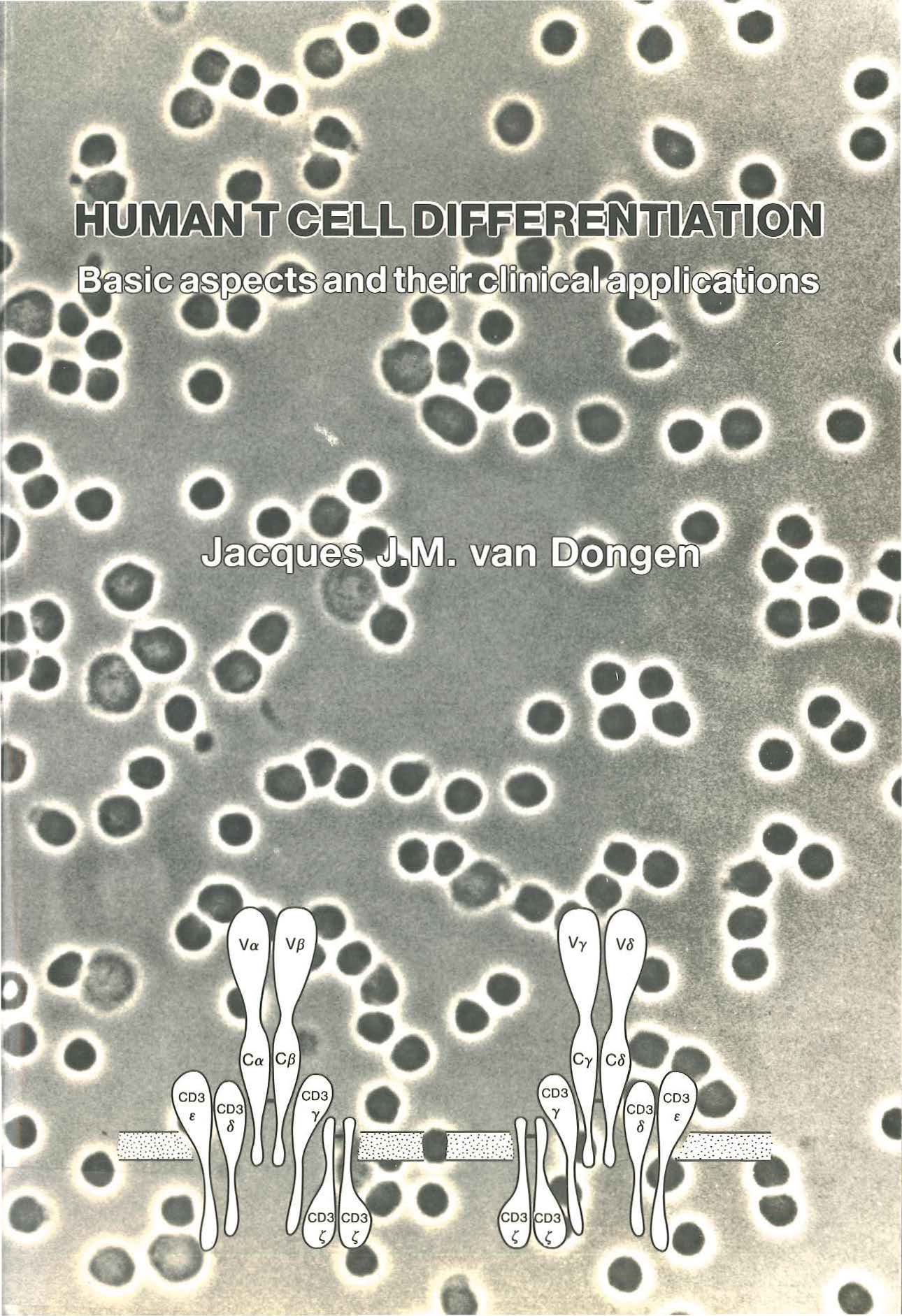
Immune recognition plays a central role in our understanding of the function of the immune system. The ability to specifically recognize foreign antigens allows selective but efficient actions of the immune system against all kinds of pathogens. This is mediated by antigen-specific receptors on B and T lymphocytes. Immunoglobulin (lg) molecules represent the antigenspecific receptors of B lymphocytes, while the T cell receptor (TeA) has this function in T lymphocytes (1). Although these two types of antigen receptors have remarkable similarities in protein structure and their encoding genes, they differ significantly in their ability to interact with antigens (1). Via their surface membrane lg (Smlg) molecules, B lymphocytes are able to recognize antigens in their native configuration either free in solution, on surfaces or on cell membranes (1). TeA molecules ofT lymphocytes can only recognize processed or degraded antigens which are physically associated with major histocompatibility complex (MHC) molecules (2,3). This TeA-mediated recognition is therefore called MHC-restricted antigen recognition (2,3). Expression of Smlg or TcA molecules by lymphocytes is acquired during lymphoid differentiation via several rearrangement processes in the lg or TcR genes (3-7). B lymphopoiesis mainly occurs in the bone marrow (8), while the thymus is thought to represent the main tissue compartment for T lymphopoiesis (9-11 ). During T cell differentiation in the thymus the T lymphocytes are "educated" for their future functions, i.e. T cells which recognize self antigens are eliminated (negative selection), while positive selection occurs for T cells which recognize foreign (non-self) antigens in association with self-MHC molecules (12-14). Upon recognition of a TeA-compatible antigen, T lymphocytes are activated, start to proliferate and exhibit their regulatory or cytotoxic functions (2). These T cell functions play a central role in the regulation of the immune system. The T lymphocytes probably coordinate immune processes via cellular interactions and lymphokines and in this way adjust and harmonize the actions of the immune system. The TeA consists of two chains, which are closely associated with the CD3 protein complex (TcA-CD3). The CD3 chains probably play an important role in signal transduction from the TeA to the cytoplasm (15). Two main types of TcA have been identified: the classical TcA-a.B and the alternative TcR-rcS (2,3,7). Data about the structure and rearrangement of the four TcR genes have become available during the last six years. The majority of these data are derived from in vivo and in vitro studies in mice (7,9). Experimental data on human T cell differentiation and human T cell function are mainly restricted to studies on thymocytes and T cell malignancies (10, 11, 16). We have used freshly obtained cell samples from healthy individuals as well as cell samples from leukemia patients and immunodeficiency patients to study human T cells and human T cell differentiation. Although most of these studies have a descriptive and inventory character, they reveal important information on the differentiation of T cells including the rearrangement and expression of TcR genes during their differentiation. Our studies also demonstrate that such basic information can be used for the development of new diagnostic tools. Optimal results in such studies can only be obtained, if a close collaboration is established between the clinical scientists and the laboratory scientists. Such a collaboration enables integration of the obtained results and evaluation of the sensitivity and specificity of newly developed diagnostic assays.
| Additional Metadata | |
|---|---|
| , , , | |
| Erasmus University Rotterdam | |
| R. Benner (Robbert) , A. Hagemeijer (Anne) | |
| hdl.handle.net/1765/50817 | |
| Organisation | Erasmus MC: University Medical Center Rotterdam |
|
van Dongen, J. (1990, June 20). Human T cell differentiation : basic aspects and their clinical applications. Retrieved from http://hdl.handle.net/1765/50817 |
|