1996-08-15
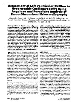
Assessment of left ventricular outflow in hypertrophic cardiomyopathy using anyplane and paraplane analysis of three-dimensional echocardiography
Publication
Publication
The American Journal of Cardiology , Volume 78 - Issue 4 p. 462- 468
This study analyzes the alterations in size and geometry of the left ventricular (LV) outflow tract that occur in hypertrophic cardiomyopathy (HC) using transthoracic 3-dimensional echocardiography. Transthoracic 3- dimensional echocardiography was performed in 17 patients with HC (4 after myectomy) and in 10 normal subjects. Images were acquired with the rotational approach, with electrocardiographic and respiratory gating. From the 3- dimensional datasets, short-axis parallel slicing of the LV outflow tract at a 1 mm distance was performed at the onset of systole. For each slice, cross- sectional area and maximal and minimal diameter were calculated. Reconstruction of the LV outflow tract could be displayed in 3 dimensions in all patients, allowing orientation and clear definition of the irregular geometry. In patients with HC, the minimal LV outflow tract cross-sectional area was smaller than in normal subjects (2.3 ± 1.0 vs 5.0 ± 0.9cm2, p <0.0001).The ratio between maximal and minimal cross-sectional areas was higher in patients with HC than in normal subjects (2.6 ± 0.9 vs 1.4 ± 0.2, p <0.0001). The ratio between maximal and minimal diameter of the smallest cross section of the LV outflow tract was also significantly higher in patients with HC than in normal subjects (1.6 ± 0.3 vs 1.2 ± 0.1, p <0.001); a value of 1.36 separated normal subjects from HC patients without previous myectomy. In conclusion, precordial 3-dimensional echocardiography allows detailed qualitative and quantitative information on the LV outflow tract. Patients with HC are characterized by a highly eccentric and asymmetric shape of the LV outflow tract, and by a smaller minimal cross- sectional area than that seen in normal subjects.
| Additional Metadata | |
|---|---|
| doi.org/10.1016/S0002-9149(96)00338-4, hdl.handle.net/1765/55602 | |
| The American Journal of Cardiology | |
| Organisation | Department of Cardiology |
|
Salustri, A., Kofflard, M., Roelandt, J., Nosir, Y., Trocino, G., Keane, D., … ten Cate, F. (1996). Assessment of left ventricular outflow in hypertrophic cardiomyopathy using anyplane and paraplane analysis of three-dimensional echocardiography. The American Journal of Cardiology, 78(4), 462–468. doi:10.1016/S0002-9149(96)00338-4 |
|