
2005-06-08
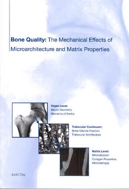
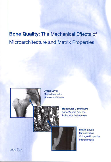
Bone Quality: The Mechanical Effects of Microarchitecture and Matrix Properties
Publication
Publication
Botkwaliteit: de mechanische effecten van microarchitectuur
In this body of work we have examined some of the current concepts pertaining to the relation between bone mass, bone quality and the mechanical properties of bone. In our first series of studies we used a model of human osteoarthritis to investigate the implications of changes in the effective tissue modulus. Having established that the material properties of the trabecular bone were altered in the earliest stages of osteoarthritis, we then investigated a possible cause, namely the breakdown or denaturation of bone collagen. Our original hypothesis was that damage at the micro scale originates at the molecular scale and that an assay of denatured collagen would refl ect the first stages of the accumulation of microdamage in bone. Although we found a significant increase in the amount of denatured collagen in early osteoarthritis, our hypothesis regarding its mechanical origin turned out to be unlikely. This points to two alternate possibilities; that either the breakdown of collagen was of an enzymatic nature, or that the quality of the original collagen was poor. In the final investigation of this series we studied the implications of a reduction of the effective tissue modulus in the presence of a normal adaptive bone response. In this study we found that when local bone strain was used as the stimulus for the mechanosensory system a reduction of the local stiffness of the bone material would result in stiffening of the bone material at the organ level. Put in a simpler way, we determined that sclerotic thickening of the subchondral bone plate could result in a stiffer plate, even if the bone material was inferior. In our second series, we investigated the effects of high dose bisphosphonate treatment in a canine model. This was part of a larger effort to quantify the effects of bisphosphonates on bone quality (150, 164, 250). Our original hypothesis was that, in addition to increasing bone mass and architectural quality, bisphosphonate treatment would result in a more mature (i.e. highly mineralized) bone matrix. This higher level of mineralization would then result in a stiffer material with improved matrix qualities. Again, we were surprised by our results! We found that any improvements in the stiffness that we could detect were due to changes in bone mass and micro-architecture alone. We also found a large, but expected, increase in the amount of microdamage present. It remains to be seen whether a similar accumulation of microdamage will occur in humans treated at clinical dosages. As is often the case in medicine, there is no easy way to improve the quality of the bone, and when not considered carefully, side effects of a treatment could be detrimental to its effi cacy. In our third series of studies we focused on the contributions of micro-architecture to bone mechanics. First we evaluated some of the morphometric tools used to quantify architecture. We found that use of the parallel plate model led to volume fraction dependant biases and recommend that direct three dimensional methods should be used whenever possible. Through the BIOMED 1 project and the Gift of Hope Organ and Tissue Donor network (not to mention the generosity of the donor’s families and the staff members who collected these specimens) Summary we acquired a unique selection of trabecular bone specimens. We used these specimens to quantify how bone architecture varied both between people and locations in the body. First, we examined the relation between bone mass and architecture. The general question that we asked could be expressed as; “If you have a certain amount of bone, in how many ways is it typically arranged?” We quantifi ed the arrangement of this bone using microCT in conjunction with standard three-dimensional morphometric measures. By using finite element models of the trabecular structure we could fully characterize the influence of microarchitecture on mechanics without considering possibly confounding matrix-level effects. In the BIOMED dataset we had samples from multiple clinically relevant sites and a wide range of donors. This provided an ideal data set to examine the effect of skeletal site. In the GOH dataset we had a large number of specimens from a small number of sites and a moderately large number of donors (this was particularly true with regards to the proximal tibia). This data set was well suited for investigating how the structure of trabecular bone varied between different people. In this analysis we found that even after correcting for the amount of bone present (BV/TV) there were indeed particular aspects of the architecture that were site specifi c. We concluded that this was probably due to differences in the mechanical function of the bone at these different sites. We also saw large differences in the bone architecture between people with equal bone mass. We could supply a striking visual representation of this by choosing 4 extreme donors from our data. For these donors, at this anatomic site, it seems that having a highly connected bone structure comes at the expense of having thin trabeculae. A small supplemental study of these four donors led us, once again, to some surprising results. Although the structure of the bone varied widely between these donors, the relation between the amount of bone present and the stiffness in the main loading direction was unaffected. It was only in the minor loading directions and in shear that the differences in architecture seemed to affect the mechanics. It had been previously demonstrated that including a morphometric measure of anisotropy improves the estimation of mechanical properties as opposed to using density alone (38, 171, 189). In our supplemental study, the differences that we observed on the minor axes as opposed to the main loading axis indicate different levels of mechanical anisotropy between these donors. If these differences could be quantified using a measure of morphometric anisotropy alone we should have been able to derive a general constitutive relation for our population i.e. by using a predictive model based on both bone mass and morphometric anisotropy we should be able to predict the mechanical properties for any donor. We tested this hypothesis by evaluating 3 different relations between the morphology and the bone’s elastic properties. Although adding anisotropy resulted in strong increases in the predictive power of the models, there were still strong site-dependant and donor-dependant differences. Unsatisfied with this result we decided to extend the one of the current models to include additional morphometric parameters. A principal components analysis demonstrated the morphology could generally be described by 3 components; one related to bone mass, one to connectivity and the last to anisotropy. We found that by including parameters such as trabecular spacing or connectivity density in the model we could improve the prediction of the model by about 20% and eliminate much of the residual error that was associated with donor and anatomic site. Although the observed improvement in predicting the mechanical properties was small, it was sufficient to validate the concept that microarchitecture does indeed influence bone mechanics. Concluding this portion of the thesis, we have demonstrated that inter-site and inter-individual differences exist in bone quality as measured by trabecular micro-architecture and that these differences can be, for a large part, quantified using existing morphometric parameters.
| Additional Metadata | |
|---|---|
| Erasmus University Rotterdam | |
| hdl.handle.net/1765/6761 | |
| Organisation | Erasmus MC: University Medical Center Rotterdam |
|
Day, J. (2005, June 8). Bone Quality: The Mechanical Effects of Microarchitecture and Matrix Properties. Retrieved from http://hdl.handle.net/1765/6761 |
|