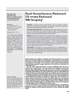
PURPOSE: To assess endoanal ultrasonography (US) and endoanal magnetic resonance (MR) imaging for mapping of anal sphincter defects that have been validated at surgery in patients with fecal incontinence. MATERIALS AND METHODS: US, MR imaging, and surgical findings in 22 women with fecal incontinence who underwent sphincter repair were retrospectively reviewed. US and MR imaging had been performed before surgery. The findings were evaluated separately and validated with surgical results. RESULTS: Endoanal MR imaging findings showed better agreement with surgical results than did endoanal US findings for diagnosis of lesions of the external sphincter (kappa value, 0.85 vs 0.53) and of the internal sphincter (kappa value, 0.64 vs 0.49). Endoanal US could not accurately demonstrate thinning of the external sphincter. MR imaging results correlated moderately with US results (kappa = 0.39). If endoanal MR images alone had been considered, the correct surgical decision would have been made in 21 (95%) patients; if endoanal US images alone had been considered, the correct decision would have been made in 17 (77%) patients. CONCLUSION: MR imaging is more accurate than US for demonstration of sphincter lesions. MR imaging provides higher spatial resolution and better inherent image contrast for lesion characterization. Endoanal MR imaging allows more precise description of the extent and structure of complex lesions and is superior for help in decisions about optimal therapy.
| Additional Metadata | |
|---|---|
| , , , , , , , , | |
| hdl.handle.net/1765/9143 | |
| Radiology | |
| Organisation | Erasmus MC: University Medical Center Rotterdam |
|
Rociu, E., Stoker, J., Eijkemans, R., Schouten, R., & Lameris, J. S. (1999). Fecal incontinence: endoanal US versus endoanal MR imaging. Radiology. Retrieved from http://hdl.handle.net/1765/9143 |
|