1987
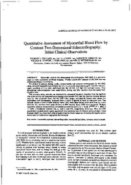
Quantitative assessment of myocardial blood flow by contrast two-dimensional echocardiography: initial clinical observations
Publication
Publication
American Journal of Physiologic Imaging , Volume 2 p. 56- 60
Myocardial contrast two-dimensional echocardiography (MC-2DE) is a new technique to study myocardial perfusion imaging. Whether quantitative analysis of MC-2DE has any clinical significance is not known. We studied 12 patients during cardiac catheterization and coronary arteriography by MC-2DE, using sonicated iopamidol (microbubble size 12 +/- 4 micron) as the echocontrast agent. Selective intracoronary injections of 4 cc were performed into the left and right coronary artery. Two-dimensional echocardiograms were made before, during, and after injection from the apical four-chamber view. The coronary artery stenosis was calculated by automated boundary detection from the digitized cine arteriograms and expressed as percentage area stenosis (%S); also the absolute minimal luminal area (L) was calculated. From the MC-2DE video images, end-diastolic frames were chosen for digitization and videointensity measured from a region of interest at basal or midseptal level. This analysis reveals a curve of echo intensity versus time. From these curves, total curve area (A), curve duration (T), and time from peak intensity to 50% intensity decay (T50) were measured. Multiple regression analysis reveals the best correlation between %S and A (A = 52.48. e0.02%S; P less than .0001; r = 0.89). Correlations between %S, L, and T and T50, respectively, were less. Thus MC-2DE quantitative analysis shows a good agreement with anatomical size of coronary artery stenosis. These findings might have important clinical implications for future follow-up of various therapeutic procedures such as transluminal angioplasty thrombolysis.
| Additional Metadata | |
|---|---|
| , , , , , , , , , , , , | |
| hdl.handle.net/1765/4238 | |
| American Journal of Physiologic Imaging | |
| Organisation | Erasmus MC: University Medical Center Rotterdam |
|
ten Cate, F., Cornel, J., Serruys, P., Vletter, W., Mittertreiner, W. H., & Roelandt, J. (1987). Quantitative assessment of myocardial blood flow by contrast two-dimensional echocardiography: initial clinical observations. American Journal of Physiologic Imaging, 2, 56–60. Retrieved from http://hdl.handle.net/1765/4238 |
|