2016-11-01
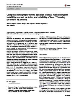
Computed tomography for the detection of distal radioulnar joint instability: normal variation and reliability of four CT scoring systems in 46 patients
Publication
Publication
Skeletal Radiology , Volume 45 - Issue 11 p. 1487- 1493
Objectives: The diagnosis of distal radioulnar joint (DRUJ) instability is clinically challenging. Computed tomography (CT) may aid in the diagnosis, but the reliability and normal variation for DRUJ translation on CT have not been established in detail. The aim of this study was to evaluate inter- and intraobserver agreement and normal ranges of CT scoring methods for determination of DRUJ translation in both posttraumatic and uninjured wrists. Materials and methods: Patients with a conservatively treated, unilateral distal radius fracture were included. CT scans of both wrists were evaluated independently, by two readers using the radioulnar line method, subluxation ratio method, epicenter method and radioulnar ratio method. The inter- and intraobserver agreement was assessed and normal values were determined based on the uninjured wrists. Results: Ninety-two wrist CTs (mean age: 56.5Â years, SD: 17.0, mean follow-up 4.2Â years, SD: 0.5) were evaluated. Interobserver agreement was best for the epicenter method [ICC = 0.73, 95Â % confidence interval (CI) 0.65–0.79]. Intraobserver agreement was almost perfect for the radioulnar line method (ICC = 0.82, 95Â % CI 0.77–0.87). Each method showed a wide normal range for normal DRUJ translation. Normal range for the epicenter method is −0.35 to −0.06 in pronation and −0.11 to 0.19 in supination. Conclusion: DRUJ translation on CT in pro- and supination can be reliably evaluated in both normal and posttraumatic wrists, however with large normal variation. The epicenter method seems the most reliable. Scanning of both wrists might be helpful to prevent the radiological overdiagnosis of instability.
| Additional Metadata | |
|---|---|
| , , , , | |
| doi.org/10.1007/s00256-016-2455-y, hdl.handle.net/1765/103685 | |
| Surgery and Traumatology | |
| Skeletal Radiology | |
| Organisation | Department of Gynaecology & Obstetrics |
|
Wijffels, M. (Mathieu), Stomp, W. (Wouter), Krijnen, P., Reijnierse, M., & Schipper, I. (2016). Computed tomography for the detection of distal radioulnar joint instability: normal variation and reliability of four CT scoring systems in 46 patients. Skeletal Radiology, 45(11), 1487–1493. doi:10.1007/s00256-016-2455-y |
|