2018-03-01
The Value of 3D Printed Models in Understanding Acetabular Fractures
Publication
Publication
3D Printing and Additive Manufacturing , Volume 5 - Issue 1 p. 37- 45
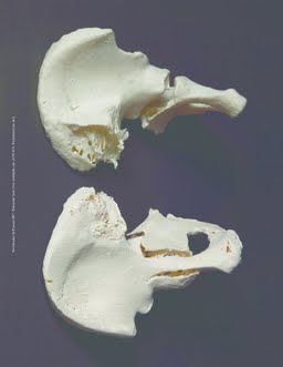
Acetabular fractures are complex and difficult to classify. Although the Judet-Letournel classification is designed to increase the understanding of acetabular fractures, it remains prone to error when using conventional medical imaging. We hypothesize that three-dimensional (3D) printing, as a new diagnostic imaging tool, will lead to an increased understanding and knowledge of acetabular fractures and an optimal surgical approach. Digital data (DICOM) of 20 acetabular fractures were converted into 3D files [standard tessellation language (STL) data]. These STL files were used to prepare 3D prints of life-size hemipelvic models with acetabular fractures. Seven senior trauma surgeons specializing in pelvic and acetabular surgery, 5 young fellowship-trained trauma surgeons, 5 senior surgical residents, 5 junior surgical residents, and 5 interns classified 20 acetabular cases using X-ray/two-dimensional (2D) computed tomography (CT), 3D reconstructions, and 3D printed models according to the Judet-Letournel classification. Furthermore, all junior and senior surgeons were instructed to evaluate their surgical approach and the positioning of the patient during operation. Time to classify each case was recorded. Calculations were done using Fleiss' κ statistics. Only slight and fair interobserver agreements for senior surgeons (κ = 0.33) and interns (κ = 0.16) were found when using X-ray/2D CT. However, 3D printed models showed moderate and substantial interobserver agreements for senior surgeons (κ = 0.59), junior surgeons (κ = 0.56), senior surgical residents (κ = 0.66), junior surgical residents (κ = 0.51), and interns (κ = 0.61). Compared with X-ray/2D CT, the interobserver agreement regarding the surgical approach for junior surgeons using 3D printed models increased by κ = 0.04 and κ = 0.23, respectively. Except for the interns, a significant time difference for classification was found between X-ray/2D CT and 3D CT and 3D printed models for junior and senior surgical residents and junior and senior surgeons (p < 0.001). 3D printing is of added value in the understanding, classification, and surgical evaluation of acetabular fractures. We recommend the implementation of 3D printed models in trauma surgery training.
| Additional Metadata | |
|---|---|
| , , , , | |
| doi.org/10.1089/3dp.2017.0043, hdl.handle.net/1765/105745 | |
| 3D Printing and Additive Manufacturing | |
| Organisation | Department of Surgery |
|
Brouwers, L. (Lars), Gunne, A.F.P.T. (Albert F. Pull Ter), de Jongh, M., van der Heijden, F., Leenen, L., Spanjersberg, W.R. (Willem R.), … Lansink, K. (2018). The Value of 3D Printed Models in Understanding Acetabular Fractures. 3D Printing and Additive Manufacturing, 5(1), 37–45. doi:10.1089/3dp.2017.0043 |
|