2017-07-20
Dynamic Organization of Chromatin Domains Revealed by Super-Resolution Live-Cell Imaging
Publication
Publication
Molecular Cell , Volume 67 - Issue 2 p. 282- 293.e7
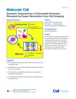
The eukaryotic genome is organized within cells as chromatin. For proper information output, higher-order chromatin structures can be regulated dynamically. How such structures form and behave in various cellular processes remains unclear. Here, by combining super-resolution imaging (photoactivated localization microscopy [PALM]) and single-nucleosome tracking, we developed a nuclear imaging system to visualize the higher-order structures along with their dynamics in live mammalian cells. We demonstrated that nucleosomes form compact domains with a peak diameter of ∼160 nm and move coherently in live cells. The heterochromatin-rich regions showed more domains and less movement. With cell differentiation, the domains became more apparent, with reduced dynamics. Furthermore, various perturbation experiments indicated that they are organized by a combination of factors, including cohesin and nucleosome-nucleosome interactions. Notably, we observed the domains during mitosis, suggesting that they act as building blocks of chromosomes and may serve as information units throughout the cell cycle.
| Additional Metadata | |
|---|---|
| , , , , , | |
| doi.org/10.1016/j.molcel.2017.06.018, hdl.handle.net/1765/108094 | |
| Molecular Cell | |
| Organisation | Erasmus MC: University Medical Center Rotterdam |
|
Nozaki, T. (Tadasu), Imai, R. (Ryosuke), Tanbo, M. (Mai), Nagashima, R. (Ryosuke), Tamura, S. (Sachiko), Tani, T. (Tomomi), … Maeshima, K. (Kazuhiro). (2017). Dynamic Organization of Chromatin Domains Revealed by Super-Resolution Live-Cell Imaging. Molecular Cell, 67(2), 282–293.e7. doi:10.1016/j.molcel.2017.06.018 |
|