2019
Cavum Septum Pellucidum in the General Pediatric Population and Its Relation to Surrounding Brain Structure Volumes, Cognitive Function, and Emotional or Behavioral Problems
Publication
Publication
American Journal of Neuroradiology , Volume 40 - Issue 2 p. 340- 346
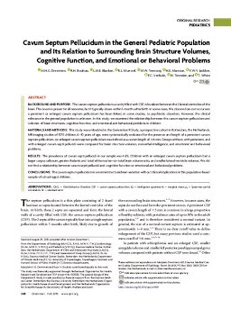
BACKGROUND AND PURPOSE: The cavum septum pellucidum, a cavity filled with CSF, is localized between the 2 lateral ventricles of the brain. The cavum is present in all neonates, but it typically closes within 5 months after birth. In some cases, this closure does not occur and a persistent or enlarged cavum septum pellucidum has been linked, in some studies, to psychiatric disorders. However, the clinical relevance in the general population is unknown. In this study, we examined the relationship between the cavum septum pellucidum and volumes of brain structures, cognitive function, and emotional and behavioral problems in children. MATERIALS AND METHODS: This study was embedded in the Generation R Study, a prospective cohort in Rotterdam, the Netherlands. MR imaging studies of 1070 children, 6 –10 years of age, were systematically evaluated for the presence and length of a persistent cavum septum pellucidum. An enlarged cavum septum pellucidum was defined as a cavum length of 6 mm. Groups without, with persistent, and with enlarged cavum septi pellucidi were compared for brain structure volumes, nonverbal intelligence, and emotional and behavioral problems. RESULTS: The prevalence of cavum septi pellucidi in our sample was 4.6%. Children with an enlarged cavum septum pellucidum had a larger corpus callosum, greater thalamic and total white matter–to–total brain volume ratio, and smaller lateral ventricle volumes. We did not find a relationship between cavum septi pellucidi and cognitive function or emotional and behavioral problems. CONCLUSIONS: The cavum septum pellucidum is a normal structural brain variation without clinical implications in this population-based sample of school-aged children.
| Additional Metadata | |
|---|---|
| doi.org/10.3174/ajnr.A5939, hdl.handle.net/1765/115227 | |
| American Journal of Neuroradiology | |
| Organisation | Department of Radiology |
|
Dremmen, M., Bouhuis, R.H., Blanken, L., Muetzel, R., Vernooij, M., El Marroun, H., … White, T. (2019). Cavum Septum Pellucidum in the General Pediatric Population and Its Relation to Surrounding Brain Structure Volumes, Cognitive Function, and Emotional or Behavioral Problems. American Journal of Neuroradiology, 40(2), 340–346. doi:10.3174/ajnr.A5939 |
|