2009-03-01
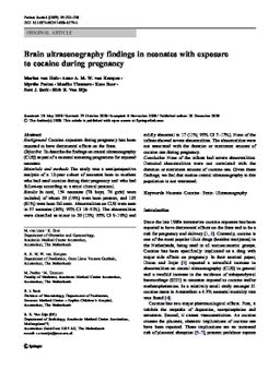
Brain ultrasonography findings in neonates with exposure to cocaine during pregnancy
Publication
Publication
Pediatric Radiology: roentgenology, nuclear medicine, ultrasonics, CT, MRI , Volume 39 - Issue 3 p. 232- 238
Background: Cocaine exposure during pregnancy has been reported to have detrimental effects on the fetus. Objective: To describe the findings on cranial ultrasonography (CUS) as part of a neonatal screening programme for exposed neonates. Materials and methods: The study was a semiprospective analysis of a 12-year cohort of neonates born to mothers who had used cocaine during their pregnancy and who had follow-up according to a strict clinical protocol. Results: In total, 154 neonates (78 boys, 76 girls) were included, of whom 29 (19%) were born preterm, and 125 (81%) were born full-term. Abnormalities on CUS were seen in 37 neonates (24%; 95% CI 18-31%). The abnormalities were classified as minor in 20 (13%; 95% CI 9-19%) and mildly abnormal in 17 (11%; 95% CI 7-17%). None of the infants showed severe abnormalities. The abnormalities were not associated with the duration or maximum amount of cocaine use during pregnancy. Conclusion: None of the infants had severe abnormalities. Detected abnormalities were not correlated with the duration or maximum amount of cocaine use. Given these findings, we feel that routine cranial ultrasonography in this population is not warranted.
| Additional Metadata | |
|---|---|
| , , , | |
| doi.org/10.1007/s00247-008-1079-3, hdl.handle.net/1765/24158 | |
| Pediatric Radiology: roentgenology, nuclear medicine, ultrasonics, CT, MRI | |
| Organisation | Erasmus MC: University Medical Center Rotterdam |
|
van Huis, M., van Kempen, A., Peelen, M., Timmers, M., Boer, K., Smit, B., & van Rijn, R. (2009). Brain ultrasonography findings in neonates with exposure to cocaine during pregnancy. Pediatric Radiology: roentgenology, nuclear medicine, ultrasonics, CT, MRI, 39(3), 232–238. doi:10.1007/s00247-008-1079-3 |
|