
1977-01-19
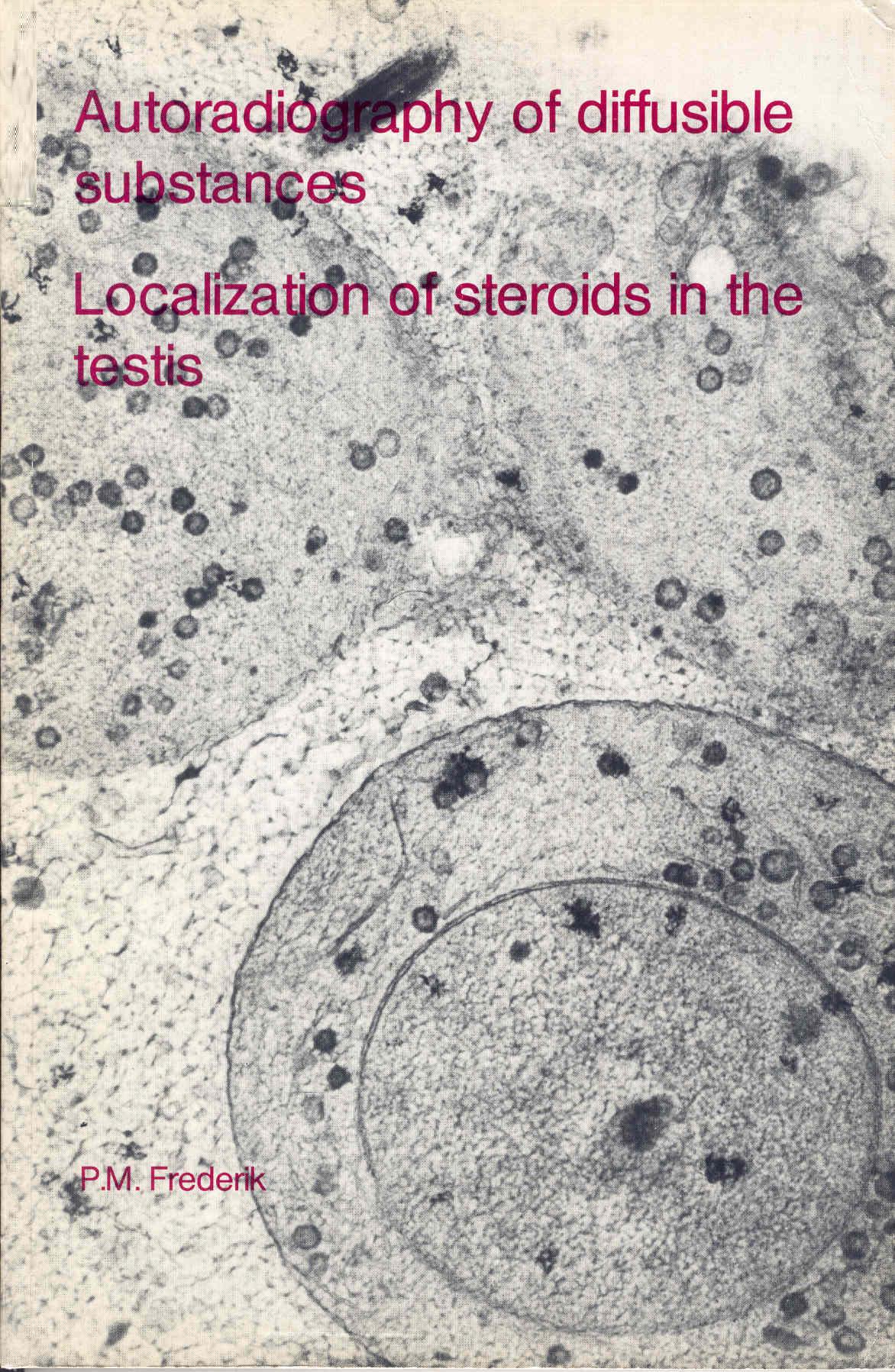
Autoradiography of diffusible substances : localization of steroids in the testis
Publication
Publication
The testis contains two cell compartments respectively having an endocrine and an exocrine function. The seminiferous tubules contain a cell renewal system concerned with the production of spermatozoa, and are the exocrine part of the organ. In the other compartment, the interstitial tissue, Leydig cells are found having an endocrine function in the biosynthesis of androgenic steroids. The studies presented in this thesis are mainly concerned with two questions regarding the role of steroids in the testis: -Firstly, which (sub) cellular structures are involved in the synthesis and secretion of androgen in the Leydig cells. -Secondly, which testicular cells or cell organelles are targets for androgen and estrogen action and what" is the route by which these steroids reach their targets. To answer these questions tritiated steroids have been administered to the rat testis under various experimental conditions. The incorporated radioactivity was subsequently localized in tissue sections by autoradiography both at the level of the light and of the electron microscope. Before discussing the merits and the technical problems of this experimental approach an outline of the role of steroids (among other factors) in testis in general and for spermatogenesis in particular will be given.
| Additional Metadata | |
|---|---|
| , | |
| Erasmus University Rotterdam | |
| hdl.handle.net/1765/25997 | |
| Organisation | Erasmus MC: University Medical Center Rotterdam |
|
Frederik, P. M. (1977). Autoradiography of diffusible substances : localization of steroids in the testis.http://hdl.handle.net/1765/25997 |
|
