2007-10-01
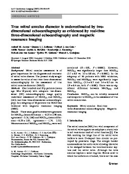
True mitral annulus diameter is underestimated by two-dimensional echocardiography as evidenced by real-time three-dimensional echocardiography and magnetic resonance imaging
Publication
Publication
International Journal of Cardiovascular Imaging , Volume 23 - Issue 5 p. 541- 547
Background: Mitral annulus assessment is of great importance for the diagnosis and treatment of mitral valve disease. The present study sought to assess the value of real-time three-dimensional echocardiography for the assessment of true mitral annulus diameter (MAD). Methods: One hundred and fifty patients (mean age 38 ± 18 years) with adequate two-dimensional (2D) echocardiographic image quality underwent assessment of MAD2Dand MAD3D(with real-time three-dimensional echocardiography). In a subgroup of 30 patients true MAD was validated with magnetic resonance imaging (MRI). Results: There was a good interobserver agreement for MAD2D(mean difference = -0.25 ± 2.90 mm, agreement: -3.16, 2.66) and MAD3D(mean difference = 0.29 ± 2.03, agreement = -1.74, 2.32). Measurements of MAD2Dand MAD3Dwere well correlated (R = 0.81, P < 0.0001). However, MAD3Dwas significantly larger than MAD2D(3.7 ± 0.9 vs. 3.3 ± 0.8 cm, P < 0.0001). In the subgroup of 30 patients with MRI validation, MAD3Dand MADMRIwere significantly larger than MAD2D(3.3 ± 0.5 and 3.4 ± 0.5 cm vs. 2.9 ± 0.4 cm, both P < 0.001). There was no significant difference between MADMRIand MAD3D. Conclusion: MAD3Dcan be reliably measured and is superior to MAD2Din the assessment of true mitral annular size.
| Additional Metadata | |
|---|---|
| , , | |
| doi.org/10.1007/s10554-006-9181-9, hdl.handle.net/1765/36970 | |
| International Journal of Cardiovascular Imaging | |
| Organisation | Erasmus MC: University Medical Center Rotterdam |
|
Anwar, A., Soliman, O. I. I., ten Cate, F., Nemes, A., Vletter-McGhie, J., Krenning, B., … Geleijnse, M. (2007). True mitral annulus diameter is underestimated by two-dimensional echocardiography as evidenced by real-time three-dimensional echocardiography and magnetic resonance imaging. International Journal of Cardiovascular Imaging, 23(5), 541–547. doi:10.1007/s10554-006-9181-9 |
|