2006
Digitisation and 3D reconstruction of 30 year old microscopic sections of human embryo, foetus and orbit
Publication
Publication
Lecture Notes in Computer Science (including subseries Lecture Notes in Artificial Intelligence and Lecture Notes in Bioinformatics) , Volume 4142 p. 636- 647
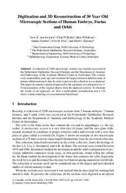
A collection of 2200 microscopic sections was recently recovered at the Netherlands Ophthalmic Research Institute and the Department of Anatomy and Embryology of the Academic Medical Centre in Amsterdam. The sections were created thirty years ago and constitute the largest and most detailed study of human orbital anatomy to date. In order to preserve the collection, it was digitised. This paper documents a practical approach to the automatic reconstruction of a 3- D representation of the original objects from the digitised sections. To illustrate the results of our approach, we show a multi-planar reconstruction and a 3-D direct volume rendering of a reconstructed foetal head.
| Additional Metadata | |
|---|---|
| , , | |
| hdl.handle.net/1765/40979 | |
| Lecture Notes in Computer Science (including subseries Lecture Notes in Artificial Intelligence and Lecture Notes in Bioinformatics) | |
| Organisation | Erasmus MC: University Medical Center Rotterdam |
|
van Zwieten, J., Botha, C., Willekens, B., Schutte, S., Post, F., & Simonsz, H. (2006). Digitisation and 3D reconstruction of 30 year old microscopic sections of human embryo, foetus and orbit. Lecture Notes in Computer Science (including subseries Lecture Notes in Artificial Intelligence and Lecture Notes in Bioinformatics), 4142, 636–647. Retrieved from http://hdl.handle.net/1765/40979 |
|