1977
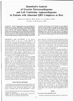
Quantitative analysis of exercise electrocardiograms and left ventricular angiograms in patients with abnormal QRS complexes at rest
Publication
Publication
Circulation (Baltimore) Issue 55 p. 55- 60
The ECG changes during exercise are described in 71 patients with a previous anteroseptal or anterolateral infarction (ANT-MI) and in 73 patients with an old posterior or inferior wall infarction (INF-MI). Left ventricular angiograms in 95 patients yielded a good correlation between areas of dyssynergy and the QRS pattern at rest. The ST changes in patients with coronary artery disease and a normal ECG at rest, and in normal subjects, were oriented toward the right, posteriorly and superiorly. In patients with INF-MI and inferior wall dyssynergy, the ST changes were more inferiorly oriented. Anteriorly-oriented ST changes were associated with anterior wall or apical dyssynergy and with ANT-MI. Thus the spatial direction of the ST changes during exercise is related to three independent factors: those factors which cause the ST changes in normal subjects, the degree of myocardial ischemia in that particular case, and the extent of dyssynergic areas in the wall of the left ventricle.
| Additional Metadata | |
|---|---|
| , , , , , , , , | |
| hdl.handle.net/1765/5223 | |
| Circulation (Baltimore) | |
| Organisation | Erasmus MC: University Medical Center Rotterdam |
|
Simoons, M., & Hugenholtz, P. (1977). Quantitative analysis of exercise electrocardiograms and left ventricular angiograms in patients with abnormal QRS complexes at rest. Circulation (Baltimore), (55), 55–60. Retrieved from http://hdl.handle.net/1765/5223 |
|