2012-05-01
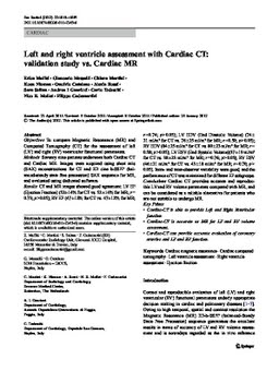
Left and right ventricle assessment with Cardiac CT: Validation study vs. Cardiac MR
Publication
Publication
European Radiology: journal of the European Congress of Radiology , Volume 22 - Issue 5 p. 1041- 1049
Objectives To compare Magnetic Resonance (MR) and Computed Tomography (CT) for the assessment of left (LV) and right (RV) ventricular functional parameters. Methods Seventy nine patients underwent both Cardiac CT and Cardiac MR. Images were acquired using short axis (SAX) reconstructions for CT and 2D cine b-SSFP (balanced- steady state free precession) SAX sequence for MR, and evaluated using dedicated software. Results CT and MR images showed good agreement: LV EF (Ejection Fraction) (52±14% for CT vs. 52±14% for MR; r0 0.73; p>0.05); RV EF (47±12% for CT vs. 47±12% for MR; r00.74; p>0.05); LV EDV (End Diastolic Volume) (74± 21 ml/m 2 for CT vs. 76±25 ml/m 2 for MR; r00.59; p>0.05); RV EDV (84±25 ml/m 2 for CT vs. 80±23 ml/m 2 for MR; r0 0.58; p>0.05); LV ESV (End Systolic Volume)(37±19 ml/m 2 for CT vs. 38±23 ml/m 2 for MR; r00.76; p>0.05); RV ESV (46±21 ml/m 2 for CT vs. 43±18 ml/m 2 for MR; r00.70; p>0.05). Intra- and inter-observer variability were good, and the performance of CT was maintained for different EF subgroups. Conclusions Cardiac CT provides accurate and reproducible LVand RV volume parameters compared with MR, and can be considered as a reliable alternative for patients who are not suitable to undergo MR. Key Points • Cardiac-CT is able to provide Left and Right Ventricular function. • Cardiac-CT is accurate as MR for LV and RV volume assessment. • Cardiac-CT can provide accurate evaluation of coronary arteries and LV and RV function.
| Additional Metadata | |
|---|---|
| , , , , | |
| doi.org/10.1007/s00330-011-2345-6, hdl.handle.net/1765/63796 | |
| European Radiology: journal of the European Congress of Radiology | |
| Organisation | Department of Cardiology |
|
Maffei, E., Messalli, G., Martini, C., Nieman, K., Catalano, O., Rossi, A., Seitun, S., Guaricci, A., Tedeschi, C., Mollet, N.& Cademartiri, F. (2012). Left and right ventricle assessment with Cardiac CT: Validation study vs. Cardiac MR. European Radiology: Journal of the European Congress of Radiology, 22(5), 1041–1049.https://doi.org/10.1007/s00330-011-2345-6 |
|