1995
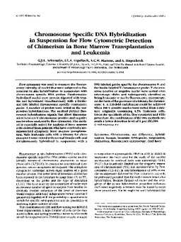
Chromosome specific DNA hybridization in suspension for flow cytometric detection of chimerism in bone marrow transplantation and leukemia
Publication
Publication
Cytometry , Volume 19 - Issue 4 p. 353- 360
Flow cytometry was used to measure the fluorescence intensity of nuclei that were subjected to fluorescent in situ hybridization in suspension with chromosome specific DNA probes. Paraformaldehyde-fixed nuclei were protein digested with trypsin and hybridized simultaneously with a biotin-and DIG labeled chromosome specific centromere probe. A number of probes were tested in the suspension hybridizations. The method yielded fluorescent hybridization signals that allow discrimination between Y chromosome positive and negative nuclei when analyzed by flow cytometry. The method is especially suited for analysis of bone marrow cells derived from patients who have received a sex-mismatched allogeneic bone marrow transplantation. Male leukemia cells with a trisomy for chromosome 8 were mixed with normal female cells and simultaneously hybridized in suspension with a DIG labeled probe specific for chromosome 8 and the biotin labeled Y chromosome probe. Y chromosome positive or negative nuclei were sorted onto microscope slides and subsequently classified as being leukemic or not by fluorescence microscopy, on the basis of the presence of a trisomy for chromosome 8. A 120-fold enrichment could be achieved when 300 Y positive nuclei were sorted from a mixture originally containing 0.5% leukemia cells. Given the specificity of the flow cytometry and FISH procedure, the combination of the two methods can reach a lower detection level of 1 per 250,000.
| Additional Metadata | |
|---|---|
| , , , , , , , , , | |
| doi.org/10.1002/cyto.990190410, hdl.handle.net/1765/67600 | |
| Cytometry | |
| Organisation | Department of Hematology |
|
Arkesteijn, G., Erpelinck, S., Martens, A., & Hagenbeek, A. (1995). Chromosome specific DNA hybridization in suspension for flow cytometric detection of chimerism in bone marrow transplantation and leukemia. Cytometry, 19(4), 353–360. doi:10.1002/cyto.990190410 |
|