1995
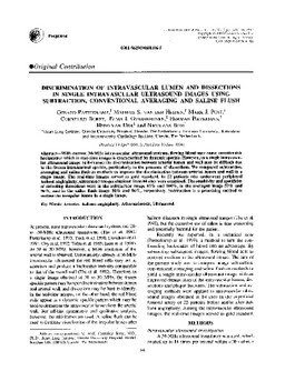
Discrimination of intravascular lumen and dissections in single intravascular ultrasound images using subtraction, conventional averaging and saline flush
Publication
Publication
Ultrasound in Medicine & Biology , Volume 21 - Issue 2 p. 149- 156
With current 30-MHz intravascular ultrasound systems, flowing blood may cause considerable backscatter which in real-time images is characterized by dynamic speckle. However, in a single intravascular ultrasound image (still-frame) the discrimination between arterial lumen and wall may be difficult due to the frozen intraluminal speckle, particularly in the presence of dissections. We compared subtraction, averaging and saline flush as methods to improve the discrimination between arterial lumen and wall in a single image. The real-time images served as gold standard. In 22 patients who underwent peripheral balloon angioplasty, ultrasound images obtained from 84 sites were examined. The sensitivity and specificity of detecting dissections were in the subtraction image 85% and 100%, in the averaged image 57% and 96%, and in the saline flush image 58% and 86%, respectively. Subtraction is a promising method to outline the irregular lumen in a single image.
| Additional Metadata | |
|---|---|
| , , , | |
| doi.org/10.1016/S0301-5629(94)00106-5, hdl.handle.net/1765/72284 | |
| Ultrasound in Medicine & Biology | |
| Organisation | Department of Surgery |
|
Pasterkamp, G., van der Heiden, M., Post, M., Borst, C., Gussenhoven, E., Pieterman, H., … Bom, K. (1995). Discrimination of intravascular lumen and dissections in single intravascular ultrasound images using subtraction, conventional averaging and saline flush. Ultrasound in Medicine & Biology, 21(2), 149–156. doi:10.1016/S0301-5629(94)00106-5 |
|