
2005-02-16
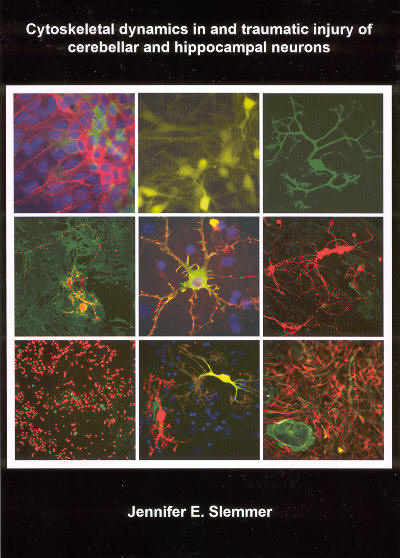
Cytoskeletal dynamics in and traumatic injury of cerebellar and hippocampal neurons
Publication
Publication
Cytoske1ete1e dynamiek en traumatische 1etse1 van cerebellaire en hippocampa1e zenuwcellen
Abstract
This thesis addresses two separate, yet overlapping, physiological processes, namely traumatic brain injury (TBI) and microtubule (MT) dynamics, primarily through the use of cultured cells derived from embryonic mice. The difficulties which arise through the experimental study of these processes, either in brain slices or in the whole animal, can be partially circumvented by using various in vitro systems to tease out the factors contributing to, and the mechanisms behind, these complex phenomena. Here, focus is given to cell cultures derived from two different brain regions, the hippocampus and the cerebellum, reviewed in this Chapter. In addition, the Introduction describes cell culture protocols, and neuronal cytoarchitecture in the hippocampus and the cerebellum, before relating these findings to glutamate-mediated cell death, particularly in cerebellar Purkinje cells (PCs). The effects of "excitotoxicity" are identified in these cells at the level of the dendritic spine and the MTs of the neuronal cytoskeleton. Alterations in both cultured PCs and hippocampal neurons are also described in relation to traumatic injury and to Williams Syndrome (WS), a rare genetic disorder. This initial portion provides a theoretical framework for the experimental studies contained in the successive chapters. In Chapter 2, cultured cells are injured using an in vitro model of stretch-induced injury. With this model, the cellular sequelae of trauma are examined in hippocampal and cerebellar cells. Several questions are asked: 1) What are the effects of increasing degrees of stretch on these cells? 2) What are the similarities and differences between neurons and glia in response to stretch injury? 3) What are the similarities and differences between hippocampal and cerebellar cells in response to stretch injury? 4) What is the relationship between TBI and glutamate-mediated excitotoxicity? and 5) What are the potential correlates among stretch injury in vitro, experimental injury to animals in vivo, and clinical TBI? Chapter 3 is concerned with the activity ofMT plus-end binding proteins, namely end-binding protein 3 (EB3) and cytoplasmic linker protein of 115 k.Da (CLIP-115), in neurons. First, cultured hippocampal and cerebellar cells are made to express EB3 tagged with green fluorescent protein (GFP) in order to image and quantify the activity of MTs in neurons and glia. Second, hippocampal brain slices from adult CLIP-115 homozygous (knockout, or KO) mice are examined in order to quantify the cellular effects of this genetic deletion. Several questions are asked: 1) What is the role of plus-end binding proteins in neurons, especially as compared to glia or to other non-neuronal cells? 2) What are the similarities and differences in the velocities of growing MTs in hippocampal neurons and glia, and in cerebellar PCs and glia? 3) What happens to MT growth velocities, and other dynamic processes, when cultured cells derived from CLIP-115 KO mice are made to express EB3-GFP? 4) What are the effects of the deletion of CLIP-115 on dendritic and spine morphologies in the adult mouse hippocampus? and 5) What are the implications of altered MT dynamics for patients with WS? Lastly, Chapter 4 presents concluding remarks and implications of the present data. Here future studies are highlighted that utilize in vitro preparations for the combined study of cytoskeletal dynamics and traumatic injury.
| Additional Metadata | |
|---|---|
| , , , | |
| C.I. de Zeeuw (Chris) | |
| Erasmus University Rotterdam | |
| hdl.handle.net/1765/76068 | |
| Organisation | Erasmus MC: University Medical Center Rotterdam |
|
Slemmer, J. (2005, February 16). Cytoskeletal dynamics in and traumatic injury of cerebellar and hippocampal neurons. Retrieved from http://hdl.handle.net/1765/76068 |
|
