2001-01-14
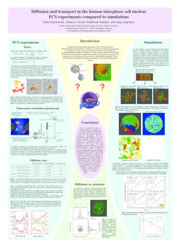
Diffusion and transport in the human interphase cell nucleus - FCS experiments compared to simulations.
Publication
Publication
Despite the succesful linear sequencing of the human genome the three-dimensional arrangement of chromatin,
functional, and structural components is still largely unknown. Molecular transport and diffusion are important
for processes like gene regulation, replication, or repair and are vitally influenced by the structure. With a
comparison between fluorescence correlation spectroscopy (FCS) experiments and simulations we show here an
interdisciplinary approach for the understanding of transport and diffusion properties in the human interphase
cell nucleus.
For a long time the interphase nucleus has been viewed as a 'spaghetti soup' of DNA without much internal
structure, except during cell division. Only recently has it become apparent that chromosomes occupy distinct
'territories' also in interphase. Two models for the detailed folding of the 30 nm chromatin fibre within these
territories are under debate: In the Random-Walk/Giant-Loop-model big loops of 3 to 5 Mbp are attached to a
non-DNA backbone. In the Multi-Loop-Subcompartment (MLS) model loops of around 120 kbp are forming
rosettes which are also interconnected by the chromatin fibre. Here we show with a comparison between
simulations and experiments an interdisciplinary approach leading to a determination of the three-dimensional
organization of the human genome: For the predictions of experiments various models of human interphase
chromosomes and the whole cell nucleus were simulated with Monte Carlo and Brownian Dynamics methods.
Only the MLS-model leads to the formation of non-overlapping chromosome territories and distinct functional
and dynamic subcompartments in agreement with experiments. Fluorescence in situ hybridization is used for the
specific marking of chromosome arms and pairs of small chromosomal DNA regions. The labelling is visualized
with confocal laser scanning microscopy followed by image reconstruction procedures. Chromosome arms show
only small overlap and globular substructures as predicted by the MLS-model. The spatial distances between
pairs of genomic markers as function of their genomic separation result in a MLS-model with loop and linker
sizes around 126 kbp. With the development of GFP-fusion-proteins it is possible to study the chromatin
distribution and dynamics resulting from cell cycle, treatment by chemicals or radiation in vivo. The chromatin
distributions are similar to those found in the simulation of whole cell nuclei of the MLS-model. Fractal analysis
is especially suited to quantify the unordered and non-euclidean chromatin distribution of the nucleus. The
dynamic behaviour of the chromatin structure and the diffusion of particles in the nucleus are also closely
connected to the fractal dimension. Fractal analysis of the simulations reveal the multi-fractality of
chromosomes. First fractal analysis of chromatin distributions in vivo result in significant differences for
different morphologies and might favour a MLS-model-like chromatin distribution. Simulations of fragment
distributions based on double strand breakage after carbon-ion irradiation differ in different models. Here again a
comparison with experiments favours a MLS-model.
FCS in combination with a scanning device is a suitable tool to study the diffusion characteristics of fluorescent
proteins in living cell nuclei with high spatial resolution. Computer simulations of the three-dimensional
organization of the human interphase nucleus allows a detailed test of theoretical models in comparison to
experiments. Diffusion and transport in the nucleus are most appropriately described with the concept of
obstructed diffusion. A large volume fraction of the nucleus seems to contain a cytosol-like liquid with an
apparent viscosity 5 times higher than in water. The geometry of particles and structure as well as their
interactions influence the mobilities in terms of speed and spatial coverage. A considerable amount of genomic
sites is accessible for not too large particles. FCS experiments and simulations based on the polymer model are
in a good agreement. Using recently developed in vivo chromatin markers, a detailed study of mobility vs.
structure is subject of current work.
| Additional Metadata | |
|---|---|
| , , , , , , , , , , , , , , , , , , , , , , , , , , , , , , , , , , , , , , , , , , , , , , , , , , , , , , , , , , , , , , , , , , , , , , , , , , , , , , , , | |
| hdl.handle.net/1765/77486 | |
| Scientific Studies from Diploma- and PhD- Students of the German Cancer Research Centre (DKFZ) | |
| Organisation | Biophysical Genomics, Department Cell Biology & Genetics |
|
Wachsmuth, M., Knoch, T., Münkel, C., & Langowski, J. (2001). Diffusion and transport in the human interphase cell nucleus - FCS experiments compared to simulations.. Presented at the Scientific Studies from Diploma- and PhD- Students of the German Cancer Research Centre (DKFZ). Retrieved from http://hdl.handle.net/1765/77486 |
|