
2008-09-17
Approaching the three-dimensional organization and dynamics of the human genome
Publication
Publication
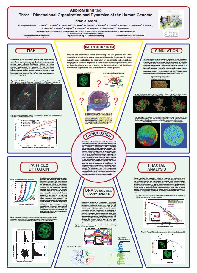
To approach the three-dimensional organization of the human cell nucleus, the structural-, scaling- and dynamic properties of interphase chromosomes and cell nuclei were simulated with Monte Carlo and Brownian Dynamics methods. The 30 nm chromatin fibre was folded according to the Multi-Loop-Subcompartment (MLS) model, in which ~100 kbp loops form rosettes, connected by a linker, and the Random-Walk/Giant-Loop (RW/GL) topology, in which 1-5 Mbp loops are attached to a flexible backbone. Both the MLS and the RW/GL model form chromosome territories but only the MLS rosettes result in distinct subcompartments visible with light microscopy and low overlap of chromosomes, -arms and subcompartments. This morphology and the size of subcompartments agree with the morphology found by expression of histone auto-fluorescent protein fusions and fluorescence in situ hybridization (FISH) experiments. Even small changes of the model parameters induced significant rearrangements of the chromatin morphology. Thus, pathological diagnoses based on this morphology, are closely related to structural changes on the chromatin level. The position of interphase chromosomes depends on their metaphase location, and suggests a possible origin of current experimental findings. The chromatin density distribution of simulated confocal (CLSM) images agrees with the MLS model and with recent experiments. The scaling behaviour of the chromatin fibre topology and morphology of CLSM stacks revealed fine-structured multi-scaling behaviour in agreement with the model prediction. Review and comparison of experimental to simulated spatial distance measurements between genomic markers as function of their genomic separation also favour an MLS model with loop and linker sizes of 63 to 126 kbp. Visual inspection of the morphology reveals also big spaces allowing high accessibility to nearly every spatial location, due to the chromatin occupancy <30% and a mean mesh spacing of 29 to 82 nm for nuclei of 6 to 12 µm diameter. The simulation of diffusion agreed with this structural prediction, since the mean displacement for 10 nm sized particles of ~1 to 2 µm takes place within 10 ms. Therefore, the diffusion of biological relevant tracers is only moderately obstructed, with the degree of obstruction ranging from 2.0 to 4.0 again in experimental agreement.
| Additional Metadata | |
|---|---|
| , , , , , , , , , , , , , , , , , , , , , , , , , , , , , , , , , , , , , , , , , , , , , , , , , , , , , , , , , , , , , , , , , , , , , , , , , , , , , , , , | |
| hdl.handle.net/1765/77694 | |
| Dynamic Organization of Nuclear Function, Cold Spring Harbor Laboratory, , 17th - 22th September, 2008 | |
| Organisation | Biophysical Genomics, Department Cell Biology & Genetics |
|
Knoch, T. (2008). Approaching the three-dimensional organization and dynamics of the human genome. Presented at the Dynamic Organization of Nuclear Function, Cold Spring Harbor Laboratory, , 17th - 22th September, 2008. Retrieved from http://hdl.handle.net/1765/77694 |
|