1996
Endoanal MRI of the anal sphincter complex: correlation with cross-sectional anatomy and histology
Publication
Publication
Journal of Anatomy: molecular, cellular and experimental morphology
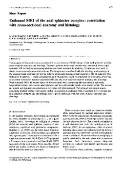
The purpose of this study was to correlate the in vivo endoanal MRI findings of the anal sphincter with the cross-sectional anatomy and histology. Fourteen patients with rectal tumours were examined with a rigid endoanal MR coil before undergoing abdominoperineal resection. In addition, 12 cadavers were used to obtain cross-sectional anatomical sections. The images were correlated with the histology and anatomy of the resected rectal specimens as well as with the cross-sectional anatomical sections of the 12 cadavers. The findings in 8 patients, 11 rectal preparations, and 10 cadavers, could be compared. In these cases, there was an excellent correlation between endoanal MRI and the cross-sectional cadaver anatomy and histology. With endoanal MRI, all muscle layers of the anal canal wall, comprising the internal anal sphincter, longitudinal muscle, the external anal sphincter and the puborectalis muscle were clearly visible. The levator ani muscle and ligamentous attachments were also well demonstrated. The perianal anatomical spaces, containing multiple septae, were clearly visible. In conclusion, endoanal MRI is excellent for visualising the anal sphincter complex and the findings show a good correlation with the cross-sectional anatomy and histology.
| Additional Metadata | |
|---|---|
| , , , , , , , , , | |
| hdl.handle.net/1765/8642 | |
| Journal of Anatomy: molecular, cellular and experimental morphology | |
| Organisation | Erasmus MC: University Medical Center Rotterdam |
|
Hussain, S., Stoker, J., Zwamborn, A. W., den Hollander, J., Kuiper, J.-W., Entius, C. A., & Lameris, J. S. (1996). Endoanal MRI of the anal sphincter complex: correlation with cross-sectional anatomy and histology. Journal of Anatomy: molecular, cellular and experimental morphology. Retrieved from http://hdl.handle.net/1765/8642 |
|