2017-05-04
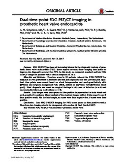
Dual-time-point FDG PET/CT imaging in prosthetic heart valve endocarditis
Publication
Publication
Journal of Nuclear Cardiology p. 1- 8
Purpose: FDG PET/CT has been of increasing interest in the diagnostic workup of prosthetic heart valve endocarditis (PVE). Some reports advocate later imaging time points to improve the diagnostic accuracy for PVE. In this study, we compared standard and late FDG PET/CT images in patients with a clinical suspicion of PVE. Materials and Methods: Fourteen scans in 13 patients referred for FDG PET/CT for suspicion of PVE performed at standard (60 min post injection) and late (150 min post injection) time points were scored based on visual interpretation and semi-quantitatively with SUVmax and target-to-background ratio (TBR, defined as [SUVmax valve/SUVmean blood pool]). Final diagnosis was based on surgical findings in all cases of infection (n = 6) and unremarkable follow-up in all others (n = 8). Results: Late images were more prone to false positive interpretation for both visual and semi-quantitative analyses. Visual analysis of the standard images yielded 1 false negative and 1 false positive result. On the late images, no scans were false negative but 5 scans were false positive. Conclusion: Late FDG PET/CT imaging for PVE seems prone to false positive results. Therefore, late imaging should be interpreted with caution.
| Additional Metadata | |
|---|---|
| , , | |
| doi.org/10.1007/s12350-017-0902-3, hdl.handle.net/1765/99746 | |
| Journal of Nuclear Cardiology | |
| Organisation | Erasmus MC: University Medical Center Rotterdam |
|
Scholtens, A. M., Swart, L., Verberne, H. J., Budde, R., & Lam, M.G.E.H. (2017). Dual-time-point FDG PET/CT imaging in prosthetic heart valve endocarditis. Journal of Nuclear Cardiology, 1–8. doi:10.1007/s12350-017-0902-3 |
|