1983
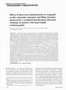
Effects of short-term administration of verapamil on left ventricular relaxation and filling dynamics measured by a combined hemodynamic-ultrasonic technique in patients with hypertrophic cardiomyopathy
Publication
Publication
Circulation (Baltimore) , Volume 68 p. 1274- 1279
The effects of short-term administration of verapamil on left ventricular isovolumetric relaxation and early and late diastolic filling dynamics were studied in 10 patients with hypertrophic cardiomyopathy by a combined hemodynamic-ultrasonic technique. Left ventricular pressures (recorded with high-fidelity micromanometers) were determined simultaneously with M mode echocardiography. After 10 mg of verapamil was given intravenously (2 mg/min), left ventricular contractility and systolic pressure dropped significantly (p less than .05). Left ventricular dP/dt fell from 1947 +/- 544 to 1489 +/- 334 mm Hg/sec, maximal velocity of the contractile element at zero load fell from 50 +/- 17 to 42 +/- 15 1/sec, peak velocity contraction of the contractile element fell from 37 +/- 10 1/sec to 29 +/- 10 1/sec (p less than .05), and left ventricular systolic pressure fell from 149 +/- 30 to 127 +/- 22 mm Hg. Left ventricular negative dP/dt increased from 1770 +/- 479 to 1477 +/- 377 mm Hg/sec (p less than .05), and the time constant of isovolumetric pressure decay was prolonged from 48 +/- 9 to 64 +/- 15 msec (p less than .05). Left ventricular end-diastolic pressure rose from 21 +/- 7 to 23 +/- 6 mm Hg (p less than .05). The time constant of isovolumetric pressure decay was calculated in three different ways, but none of these measurements was influenced by verapamil. Time of isovolumetric relaxation, duration of rapid ventricular filling, and peak rate of left ventricular lengthening were not significantly influenced by verapamil and remained highly abnormal. In contrast, peak rate of left ventricular posterior wall thinning declined further after verapamil from 2.9 +/- 1.2 to 2.4 +/- 1.4 1/sec (p less than .05).(ABSTRACT TRUNCATED AT 250 WORDS)
| Additional Metadata | |
|---|---|
| , , , , , , , , , , , , , , , | |
| hdl.handle.net/1765/4107 | |
| Circulation (Baltimore) | |
| Organisation | Erasmus MC: University Medical Center Rotterdam |
|
Serruys, P., Mey, S., Roelandt, J., & ten Cate, F. (1983). Effects of short-term administration of verapamil on left ventricular relaxation and filling dynamics measured by a combined hemodynamic-ultrasonic technique in patients with hypertrophic cardiomyopathy. Circulation (Baltimore), 68, 1274–1279. Retrieved from http://hdl.handle.net/1765/4107 |
|