
1983-03-02
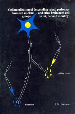
Collateralization of descending spinal pathways from red nucleus and other brainstem cell groups in rat, cat and monkey
Publication
Publication
The somatotopically organized rubrospinal pathway is the major component of the laterally descending brainstem pathways, and is especially involved in steering of fractionated movements of the distal parts of the limbs. Electrophysiological studies in cat showed that this fiber system, in contrast to the medially descending pathways, has a limited degree of collateralization in the spinal cord (Abzug et al., 1973 and 1974; Shinoda et al., 1977). The red nucleus projects also to the contralateral cerebellum (Brodal and Gogstad, 1954; Courville and Brodal, 1966). The collateralization of the rubrospinal neurons to the contralateral cerebellar interpositus nucleus is relatively high as indicated by anatomical and electrophysiological findings (Anderson, 1971; Brodal and Gogstad, 1954). Thus, the findings in these studies suggested that almost all rubrocerebellar fibers are collaterals from rubrospinal neurons. In view of the above data it appears that the rubrospinal pathway represents a focussed system, which distributes its fibers to specific groups of spinal segments. On the other hand, the rubrocerebellar pathway probably represents a direct, tightly coupled return projection to the interpositus nucleus, which is the main source of afferents to the rubrospinal neurons (Courville, 1966b; Dekker, 1981; Flumerfelt et al., 1973; King et al., 1973; Tsukahara et al., 1967). In the present anatomical study an attempt has been made to demonstrate anatomically the existence of collaterals in the rubrospinal pathway and to compare quantitatively the degree of this collateralization in rat, cat and monkey. This was done with the aid of the multiple retrograde fluorescent tracer technique. This technique will be described in chapter II. In this anatomical study one fluorescent tracer was injected in the cervical grey and another in more caudal segments of the cord. In these experiments the distribution of sin3le and double labeled neurons in red nucleus was studied. The descending pathways from the ventrolateral pontine tegmentum and from the raphe magnus, including the adjoining ventral reticular formation, also descend in the dorsolateral funiculus in rat, cat and monkey and also terminate in the dorsal grey (Basbaum and Fields, 1978, 1979; Holstege et al., 1979; Kuypers and Maisky, 1977; R.F. Martinet al., 1978; G.F. Martinet al., 1979 and 1981a; Tohyama et al., 1979a+b). Therefore, in these retrograde fluorescent double labeling studies the degree of the collateralization ~n these descending tracts was compared to that of the rubrospinal pathway ~n each of three mammalian species (i.e. rat, cat and monkey). In addition, in cat an attempt was made to determine the location of the rubro-olivary neurons in relation to the rubrospinal ones and to establish whether the rubro-olivary neurons give collaterals to the spinal cord. These studies will be presented in Chapter III and IV. In these studies attention was also paid to the rubrocerebellar connections. Specifically an attempt has been made to determine whether these connections are established by collaterals of rubrospinal neurons. These findings will be presented ~n Chapter V. Electron microscopic studies in rat, combining the anterograde and retrograde intra-axonal transport techniques, showed that many rubrospinal neurons receive afferents from the cerebellar interpositus nucleus (Dekker, 1981). Some of the fluorescent tracers used in the present study can be employed both retrogradely to demonstrate parent cell bodies as well as anterogradely to demonstrate the fiber terminals. Using the fluorescent tracers in this fashion it could be demonstrated in light microsc0py that the cerebellar interposito-rubral fibers establish contact with rubrospinal neurons. These data are also presented ~n Chapter V.
| Additional Metadata | |
|---|---|
| , , , , , | |
| Erasmus University Rotterdam | |
| H.G.J.M. Kuypers (Hans) | |
| hdl.handle.net/1765/51247 | |
| Organisation | Erasmus MC: University Medical Center Rotterdam |
|
Huisman, M. (1983, March 2). Collateralization of descending spinal pathways from red nucleus and other brainstem cell groups in rat, cat and monkey. Retrieved from http://hdl.handle.net/1765/51247 |
|